Abstract
Background. Skin incision designs for rhytidectomy have changed little since Jacques Joseph’s descriptions of the 1920s, but facial rejuvenation shifted fundamentally in 1976 with Mitz and Peyronie’s description of the superficial musculo-aponeurotic system (SMAS). Deep-layer repositioning has since focused on optimizing vector dynamics. Debate nevertheless persists regarding the ideal SMAS transposition vector—vertical versus horizontal—and the interplay between SMAS incision levels, skin redraping direction, and skin tension.
Methods. This article presents an analysis of facial aging, with particular emphasis on the structural deflation, descent, and inferomedial rotation of midface fat compartments. A focused literature review contrasts horizontal, vertical, and multi-directional vectors. Against this backdrop, the author evaluates a 40-year clinical experience using diverse SMAS modification techniques, analyzing how vector selection influences longevity, structural harmony, and the prevention of an “operated” appearance.
Results. Long-term clinical observation indicates that optimal elevation with preservation of facial harmony requires a differentiated, triple-vector approach. The most reliable outcomes result from a superolateral rotational vector for the zygomatic (malar) SMAS, a vertical vector for the parotid SMAS, and an oblique postero-superior vector applied to the platysma–SMAS complex anchored toward the mastoid eminence. This tri-directional framework decouples midface repositioning from lower-third and neck vectors, thereby relieving the skin of tension and avoiding the “windswept” appearance.
Conclusions. Surgical consensus regarding optimal rhytidectomy vectors remains non-uniform. Our long-term clinical experience suggests that delivering substantial rejuvenation while avoiding lateral distortion requires a differentiated approach: a rotational vector in the malar area, a vertical vector in the cheek, and an oblique vector in the cervical region. This combination offers the most anatomically faithful replication of youthful tissue distribution.
Introduction
The introduction of the superficial musculo-aponeurotic system (SMAS) in 1976 by Mitz and Peyronie, within Professor Paul Tessier’s department, fundamentally shifted the paradigm of facial rejuvenation (1). Manipulation of this anatomical layer allows repositioning of the deeper facial structures, including the malar fat pads, which descend and rotate inferomedially under the effects of gravity and aging. Effective repositioning requires incision and undermining of the SMAS, potentially including release of the suspensory retaining ligaments described by Furnas.
 Techniques have ranged from simple plication and suspension sutures—which involve no surgical undermining—to localized SMASectomies. Since the publication of the composite facelift by the late Dr. Sam Hamra of Dallas (2), there has been a resurgence of deep-layer techniques gathered under the “deep plane facelift” label. The most popular contemporary variant is a modification of the composite lift published by Jacono (3).
A critical question, however, has received less attention than it deserves: the differential vectors applied to the skin versus the SMAS. These vectors are not, and should not be, parallel when the goal is a stable, effective, and natural-looking rejuvenation that does not distort facial features (Figure 1).
The refinement of concepts concerning the ideal direction of SMAS vectors during a two-plane (skin + SMAS) cervicofacial facelift demands a precise anatomical understanding of this complex layer, which remains a subject of debate among anatomists (7, 8). The central premise of this article is that the SMAS is a fibrous sheet capable of tolerating multiple, non-parallel vectors for re-tensioning; one of these vectors—upward and medial rotation of the superior SMAS—is rarely made explicit in the literature.
Purpose. This article aims to clarify SMAS incision design and vector selection in cervicofacial rhytidectomy. We propose a triple-vector framework in which vector direction is differentiated by facial zone, and we describe its operative application across two complementary techniques developed over four decades of practice.
Anatomical Rationale
The malar fat pad and its center of rotation
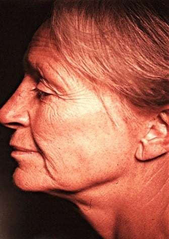
Facial aging involves structural deflation, descent, and rotation of the midface fat compartments. The submalar fat compartment undergoes a downward rotation that originates from a central paranasal point, corresponding to the vascular hilum of the emerging nasal branch of the facial artery (Figure 2). Correcting this rotation therefore requires an upward and outward (rotational) movement, not merely a vertical lift.
Rotation of the submalar fat: clinical observation and anatomical basis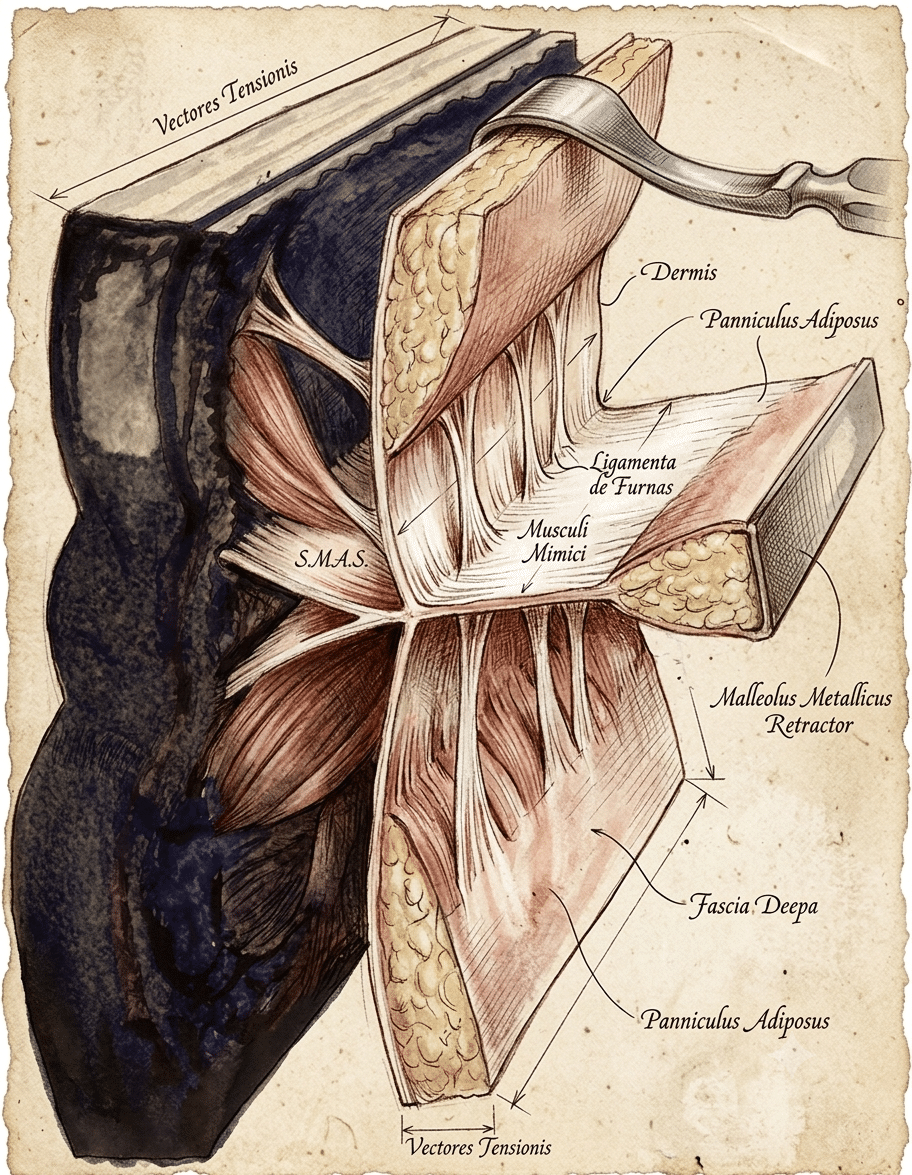
The existence of a rotation of the submalar fat compartment is a hypothesis that appears supported by examination of serial photographs of several film actors for whom images from youth and from progressively later decades are available. Thus, in Frank Sinatra and others, one observes a predominance of fat accumulation above the nasolabial fold, contemporaneous with a deflation of fat below the malar bone, involving not only the deep fat compartment but also the subcutaneous fat. This observation is further validated by examination of the author’s own face, who notes the same evolution on himself as his face has aged.
Rather than attributing this phenomenon to a simple vertical descent, it seems more accurate to posit a rotational movement whose center is paranasal—precisely where the deep vascular island lies, and which, moreover, constitutes the anatomical basis of the superior-pedicle nasolabial flaps widely used in reconstructive surgery.
The fat situated above the nasolabial fold rests on the termination of the facial SMAS; this is why it appears useful to re-tension the SMAS not only along a vertical vector, but also to combine it with a rotational movement directed upward and inward, in order to horizontalize this fat accumulation. This maneuver restores a region that we have described under the name of submalar hiatus, or mid-cheek groove.
This technical concept has been applied by the author since the 1980s, and results in an improved appearance of the cheekbone by capturing locally reflected light, thereby conferring a rejuvenated yet natural appearance to the face, without the absolute necessity of performing malar augmentation by implants or fat grafts.
The SMAS as a re-tensioning sheet
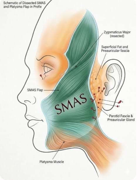
The SMAS is a surgically isolable fibrous layer, continuous with the temporalis fascia superiorly and the platysma inferiorly. It can be dissected as a substantial flap to provide an effective tension-bearing layer, provided it is not excessively thin—as occurs in some patients (Figures 4–6). Its solid parotid portion offers a reliable anchoring substrate, while the sub-zygomatic SMAS is available for elevation and rotation (Figures 6).
Redundancy, force transmission, and nerve considerations
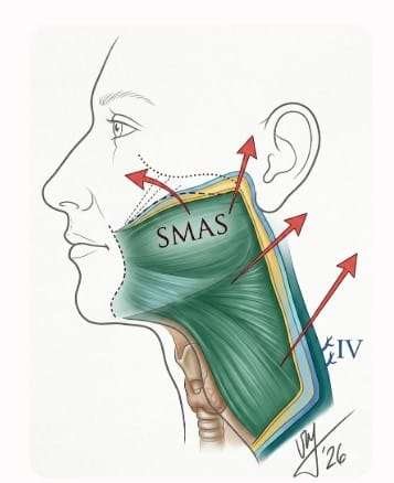 A large quadrangular SMAS flap, whose inferior section is made approximately 6–7 cm below the mandibular border, can be redraped like a hammock suspended toward the mastoid (Figures 5, 6). By tensioning this single musculo-aponeurotic (SMAS–platysma) flap, the skin is relieved of tension and simply draped—thereby avoiding a stiff or surgical (“windswept”) appearance. In selected cases, the excess SMAS resected from the upper portion of the quadrilateral can be re-implanted as viable tissue, for instance to augment the lips (Figure 3).
Dissection is straightforward in the juxta-parotid region; however, once the anterior limit of the parotid is crossed, extreme caution is required to avoid injury to the motor branches of the facial nerve, which lie immediately beneath the SMAS). Furnas retaining ligaments are separated with care and precision to maximize SMAS mobilization.
The Triple-Vector Framework
The organizing concept of this article is that SMAS re-tensioning should be differentiated by facial zone rather than applied as a single direction across the whole face. We propose three vectors (Figure 1):
- Superolateral rotational vector (malar / zygomatic region).
- Anatomy: Directed upward and backward (toward the root of the helix and the temporal region), with a rotational component around the origin of the zygomaticus major muscle. It directly addresses the vertical ptosis of the cheek and submalar soft tissues, counteracting the inferomedial rotation of the malar fat pad (Figure 2).
- Clinical implication: Firm repositioning of the SMAS flap along this axis restores midface fullness and corrects the nasolabial fold without creating a horizontal line at the oral commissure. The excess SMAS resected superiorly reflects this upward and inward rotation (Figure 3).
- Postero-superior (oblique) vector (mandibular angle and parotid rim).
- Anatomy: Oblique, directed upward and backward toward the earlobe and upper pre-auricular region.
- Clinical implication: This is the key vector for redefining the mandibular line (the “oval” of the face) and treating jowls. Its tension reshapes the cervico-mandibular angle by transmitting traction evenly along the fixed posterior rim.
- Oblique cervical vector (platysma and retro-auricular region).
- Anatomy: Directed posteriorly and superiorly, along the anterior border of the sternocleidomastoid (SCM) muscle toward the mastoid region.
- Clinical implication: This vector addresses the cervical component of the facelift. The anatomical continuity between the SMAS and the platysma allows this postero-superior traction to tighten platysmal bands, correct skin and muscle laxity of the neck, and reposition the external jugular vein in a protected, harmonious relationship.
Key principle—non-parallel vectors. Crucially, the tension vectors for the skin and the underlying SMAS are not always superimposed. This is the fundamental advantage and rationale of the bi-plane approach: by absorbing tension in the SMAS–platysma flap, the skin is freed to redrape without tension.
Personalization. The angle of these three vectors is not fixed; it must be adapted to the patient’s morphology (long and gaunt face versus short and heavy face) to restore volume without artificially widening the midface.
 Surgical Techniques
In our practice we differentiate two complementary approaches based on patient presentation and goals. The bi-plane (“Grand”) lift embodies the full triple-vector framework; the Micro SMAS lift is a limited-indication variant that applies a simplified version of the same rotational principle.
- The “Grand” Cervicofacial Lift (High SMAS / Bi-Plane)
Historically termed the “Bi-Plane” lift, this technique involves two distinct dissection planes: a wide subcutaneous undermining extending potentially to the nasolabial folds, and a deep SMAS incision in the fixed extra-parotid portion. The SMAS incision comprises three components (Figure 4):
- A primary long horizontal, sub-zygomatic incision toward the origin of the zygomaticus major muscle.
- A vertical pre-tragal incision transitioning inferiorly to incise the platysma up to 7 cm above the mandibular border, in front of the external jugular vein.
- A third horizontal counter-incision, parallel to the mandible (approximately 7 cm inferior), transecting the platysma toward the midline. The external jugular vein must be identified and preserved to avoid bleeding and hematoma.
This creates a robust, quadrangular SMAS flap (Figure 5). Although solid in most patients, it can be attenuated and less effective in others.
Dissection and vector application. After careful sub-SMAS undermining and release of the Furnas ligaments, the three vectors of the framework are applied (see above). The quadrangular flap is ideal for the superolateral rotational correction of the malar fat pad (Figure 3). The inferior portion of the SMAS–platysmal flap addresses the liberated platysma, creating a “muscular hammock” fixed to the SCM origin at the mastoid fascia. In our experience this maneuver effectively corrects submandibular gland ptosis without requiring glandular excision in most cases.
Skin vectors (distinct from SMAS vectors). Skin tension is applied along two vectors:
- A vertical vector anterior to the ear, lifting the temporal and pre-auricular skin strictly upward.
- A 45-degree (postero-superior) vector on the neck skin, anchoring skin to the mastoid region.
- The Micro SMAS Lift
The Micro SMAS lift is a composite (skin–SMAS) lift developed since 2014 and published by the author in 2018 (4). Skin and SMAS are incised and elevated together, with no or minimal section of the retaining ligaments. The SMAS incision is made in continuity with the skin incision, approximately 2–3 cm anterior to the tragus, within the thick parotid SMAS layer—much more posterior than in the deep-plane approach.
It addresses younger patients with moderate cheek ptosis, early jowls, and moderate neck flaccidity. It is offered as a surgical alternative to thread lifts and is designed to minimize post-operative swelling and social downtime. It is also an excellent option for revising previous bi-plane lifts or inefficient subcutaneous-only lifts.
Technique.
- Incision: The cutaneous incision is limited, stopping at the sideburn (Figure 7). The SMAS incision is made in continuity with the skin incision, respecting a thin fibrous tissue layer external to the parotid gland.
- Dissection: Sub-SMAS undermining is minimal, avoiding extensive disruption of the retaining ligaments. A prudent, limited dissection—usually 2–3 cm—is sufficient to mobilize the entire composite facial mask; no subperiosteal approach is required.
- Closure: The S-shaped SMAS incision allows a vest-over-pants (en paletot) suture of the liberated SMAS onto the residual SMAS anterior to the tragus—the point of maximum deep-plane tension. This S-shape also induces the necessary superior and posterior rotational vector, which repositions the malar fat pad and restores malar projection.
Clinical Experience and Results
This technical analysis is based on 40 years of surgical experience, detailed in The Art of Facelifting (Thieme, 2022). Objective comparison in rhytidectomy is notoriously difficult: patient anatomy varies, problems are not superimposable, and criteria for “improvement” are not absolute. Surgical judgment refined by experience remains the primary driver of technical evolution. The outcomes of the two procedures are not interchangeable, as their indications differ.
Efficacy and durability. The bi-plane (“Grand”) lift provides demonstrably superior, more convincing, and more durable correction of jowling, cervical laxity, and centro-facial volume (Figures 12-15. In our experience, it offers a 2- to 5-year advantage in stability over the Micro SMAS lift.
Recovery. The Micro SMAS lift remains highly effective for the appropriately selected younger patient, providing very satisfactory results with significantly reduced recovery (Figures 11, 12). Social and professional downtime is typically 5–10 days, compared with 3–6 weeks for the bi-plane lift.
Revision rate (Micro SMAS). We observed a 3% deterioration rate at one year (beginning at the 6-month post-operative mark), justifying surgical revision. One case, later diagnosed with Ehlers-Danlos syndrome, required two revisions. The jowls start to deteriorate and reappear,and sometimes the neck loses its straightness: the patient usually asks for a redo that we accomplish under strict local anesthesia, after one year post op,This secondary revision does not imply a major undermining, but consists on a very limited microSMAS lift.
Complications. Complication rates were comparable across both series:
- Minor scar infections: 2%
- Hypertrophic scarring (pre- or post-auricular): 4%
- Minor marginal necrosis: higher in bi-plane lifts (2%) versus 1 case in Micro SMAS
- Transient nerve paresis (temporal or zygomatic branch): 1%
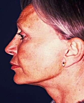
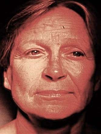
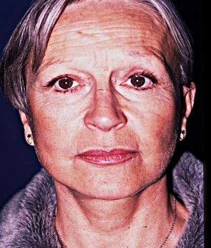


Discussion: The Vectors Debate
The manipulation of subcutaneous tissue was noted by Bourguet (5) in 1919, but the pivotal innovation came from Skoog (6). By 1972, Skoog was tensioning extra-parotid tissues using an oblique—largely horizontal—vector on the platysma. The formal surgical concept of the SMAS was then introduced by Mitz and Peyronie in 1976. Through anatomical dissection, histology, and injected cross-sections, we defined the SMAS as a surgically isolable fibrous layer, continuous with the temporalis fascia superiorly and the platysma inferiorly.
This concept was immediately controversial. Jost and Levet (7) famously debated it, and Minelli, Van der Lei, and Mendelson (8) recently (2024) questioned the existence of the SMAS as a distinct anatomical entity—controversies that persist alongside its surgical utility. Following its presentation in the United States in 1976, Owsley (9) published the first applied SMAS technique, and Connell (cited by Marten (10)) began experimenting with differential SMAS flaps. At that time, the author used the quadrangular SMAS flap described above. Years later, Hamra (2, 11) published the composite lift (1989), advocating essentially vertical skin vectors and horizontal SMAS vectors.
This history leads to the central conflict: horizontal versus vertical vectors.
The case for horizontal (or quasi-horizontal) vectors
(e.g., Skoog (6), Baker (12))
- Limited incisions and SMASectomy: A horizontal pull allows the incision to stop at the sideburn, avoiding a temporal extension. A vertical or slightly oblique SMASectomy allows efficient backward pull of the SMAS.
- Patient expectation: It mimics the “mirror test,” in which patients pull their cheeks backward horizontally to demonstrate the desired outcome.
- Drawback: This vector—especially when applied to both skin and SMAS, as in some deep-plane variants—flattens the malar region and can create a “fish-mouth” deformity by pulling the oral commissures laterally. It may also produce visible sub-dermal tension bands.
The case for vertical vectors
(e.g., Mitz, Bonnefon (13), Barton (14), Marten (10))
- Jowl correction: The vertical vector is mechanically superior for correcting jowling and lower-face ptosis, though the challenge lies in achieving resistant anchoring of the SMAS flap.
- Volumetric restoration: Most importantly, it creates an upward and inward rotation of the centro-facial and malar fat pads (Figures 6, 7). This “re-inflates” the cheekbone, restoring youthful projection and capturing light more effectively, achieving a positive volumetric effect without lipofilling in many cases.
- Challenge: The primary difficulty is identifying a solid, stable anchoring point. The long suture of the SMAS to itself below the zygomatic arch offers a reliable fixation solution.
- The rotational component: We have emphasized the rotational component of the vertical vector since the 1980s; a recent publication by Sadati et al. (15) has likewise pointed out its importance.
The synthesis: oblique (deep-plane) vectors
(e.g., Hamra (2), Jacono (16))
Synthesists use an oblique (approximately 45-degree) vector via the deep-plane approach. While the modern deep-plane lift has gained immense popularity, it remains a compromise vector.
Conclusion
The debate over rhytidectomy vectors divides practitioners into three camps: the “horizontalists” (e.g., Skoog, Baker), who prioritize a posterior pull; the “verticalists” (e.g., Mitz, Bonnefon, Barton, Marten), who prioritize a superiorly directed lift, especially of the midface; and the “synthesists” (e.g., Hamra, Jacono), who use an oblique (45-degree) vector via the deep-plane approach.
We maintain that the vertical vector—particularly in the parotid region—remains an under-utilized standard of quality for true anatomical repositioning of the malar fat. Our long-term experience further indicates that a single vector is insufficient: a triple-vector framework (rotational in the malar area, vertical in the cheek, oblique in the cervical region), with skin and SMAS vectors deliberately non-parallel, offers the most anatomically faithful replication of youthful tissue distribution.
The field urgently lacks rigorous, long-term, objective comparative studies. Until such data exist, surgical judgment and a deep understanding of anatomy—not trends—must guide the choice of vector. It remains scientifically impossible to prove that one technique is superior to another, given the variety of anatomical presentations in each individual case; meta-analyses are useful for comparing complication rates, not the quality of results. The experienced surgeon therefore follows a slow learning curve, choosing the technique most appropriate for each individual case. As Nahai has observed, there is no standard procedure in facelifting—there is no facelift for all seasons.
References
- Mitz V, Peyronie M. The superficial musculo-aponeurotic system (SMAS) in the parotid and cheek area. Plast Reconstr Surg. 1976;58:80–88.
- Hamra ST. Deep plane facelift. Paper presented at: American Society of Aesthetic Plastic Surgery Midwinter Symposium of Facial Rejuvenation; January 12–13, 1989; Santa Barbara, Calif.
- Jacono AA, Talei B. Vertical neck lifting. Facial Plast Surg Clin North Am. 2014;22(2):285–316. doi:10.1016/j.fsc.2014.01.006. PMID: 24745389.
- Mitz V. Le microlift biplan. Principes, technique, résultats. Ann Chir Plast Esthet. 2018:262–269.
- Bourguet J. La disparition chirurgicale des rides et plis du visage. Bull Acad Med (Paris). 1919.
- Skoog T. Plastic Surgery: New Methods and Refinements. Philadelphia: Saunders; 1974.
- Jost G, Levet Y. Parotid fascia and face lifting: a critical evaluation of the SMAS concept. Plast Reconstr Surg. 1984;74(1):42–51.
- Minelli L, Van der Lei B, Mendelson BC. The superficial musculoaponeurotic system: does it really exist as an anatomical entity? Plast Reconstr Surg. 2024;153(5):1023–1034.
- Owsley JQ Jr. SMAS-platysma face lift. Plast Reconstr Surg. 1983;71(4):573–576.
- Marten TJ. High SMAS facelift: combined single flap lifting of the jawline, cheek, and midface. Clin Plast Surg. 2008;35(4):569–603.
- Lemmon ML, Hamra ST. Skoog rhytidectomy: a five-year experience with 577 patients. Plast Reconstr Surg. 1980;65:283–297.
- Baker DC. Lateral SMASectomy. Plast Reconstr Surg. 1997;100(2):509–513.
- Bonnefon A. Deep vertical lift and its development regarding the central facial area and lower two-thirds of the neck. Our technique. Ann Chir Plast Esthet. 1999:609–616.
- Barton JR, Fritz E. The “high SMAS” face lift technique. Aesthet Surg J. 2002;22(5):481–487.
- Sadati K, Mathieu O, Cetrulo CL Jr, Lellouch AG. Advancements in face lift techniques: preservation face lift with a rotating pedicle flap. Plast Reconstr Surg Glob Open. 2025;13(3):e6619. doi:10.1097/GOX.0000000000006619.
- Jacono AA, Parikh SS. The minimal access deep plane extended vertical facelift. Aesthet Surg J. 2011;31(8):874–890.
Disclosure: Some drawings have been corrected with AI manipulation, and AI has assisted in the translation of this paper.